Echocardiography (also called cardiac echo or just echo) uses high-frequency sound waves to create a real-time video of your beating heart. The current American College of Cardiology/American Heart Association practice guidelines refer to the technique as the single most useful diagnostic test in the evaluation of patients with heart failure.
A cardiac echo image shows the size of the heart chambers and estimates the pressure within them; the thickness and motion of the heart wall; and the operation of the heart valves. The test can also depict the major blood vessels leading to and from the heart; show how forcefully your heart is pumping blood; and spot areas of the heart wall that have been injured by a previous heart attack or some other cause.
In people with heart failure, cardiac echo can reveal whether a person has systolic or diastolic heart failure. It also determines ejection fraction, the volume of blood pumped out of the left ventricle with each contraction.
Two-dimensional echocardiography displays a cone-shaped image of the moving heart. The addition of Doppler technology produces moving color images similar to those used in weather radar, and reveals the speed, direction, and volume of blood flow through the heart.
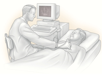
Cardiac echo is noninvasive, painless, and safe. The technician places a special wand, called a transducer, on your chest. The transducer beams ultrasound signals into your heart. The echoes of sound waves that reflect off your heart structures are translated into visual images on a computer screen.
A newer iteration, three-dimensional echocardiography, may be superior to two-dimensional in assessing size and output of the left ventricle. This technology is still evolving.